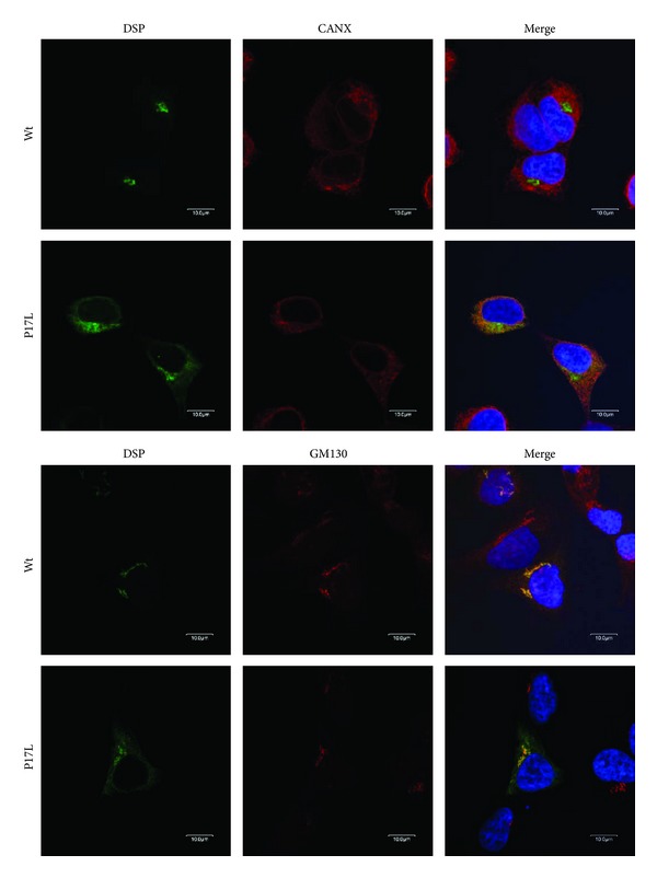
Figure 4.

Fluorescent immunocytochemistry. Confocal laser-scanning images were captured to detect localization of the GFP-tagged wildtype (Wt) and mutant (P17L) DSP in HEK293T cells. Anti-Calnexin (CANX) antibody was used for ER staining, and anti-Golgi matrix protein (GM130) antibody was used for Golgi apparatus staining. Nuclei were stained with H33342. Wildtype DSP was localized exclusively in the Golgi apparatus. The mutant DSP largely remained in the ER, although a portion was localized in the Golgi apparatus.
