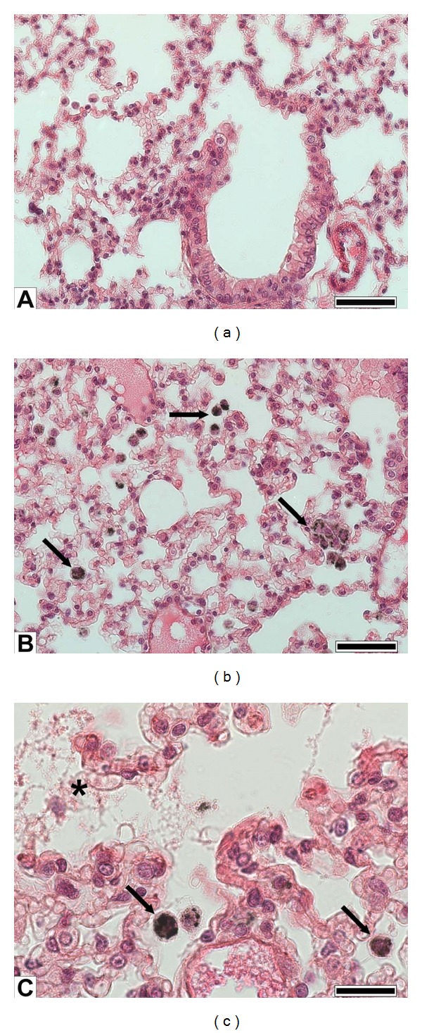
Figure 4.

Lung histology. (a) Sham lung parenchyma showing bronchiolar and alveolar epithelia; (b) PM1-treated lung showing abundant alveolar macrophages engulfing particles (arrows); (c) detail of alveoli of a lung instilled with PM1 showing particles phagocytosis by alveolar macrophages and damage of the alveolar epithelium (asterisk). (a), (b) bars = 50 μm; and (c) bar = 20 μm.
