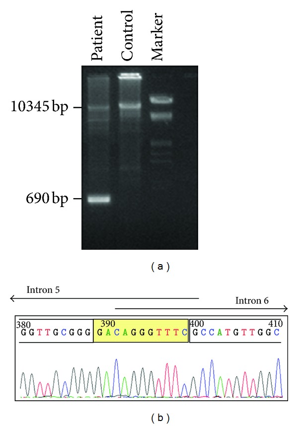
Figure 3.

Confirmation and characterisation of the MSH2 exon 6 deletion. (a) Agarose gel electrophoresis (1.5%) of the long-range PCR product obtained using the forward primer located in exon 5 (5FP) and the reverse primers located in intron 6 (6RPg) (as described in the text); DNA Molecular Weight Marker III (Roche) used. An abnormal 690 bp fragment was obtained for our patient. (b) Sequence analysis of the truncated 690-bp PCR amplicon reveals the loss of a 9,655-bp genomic region. The breakpoints highlighted in yellow are located in a strech of 11 nucleotides common to both introns 5 and 6.
