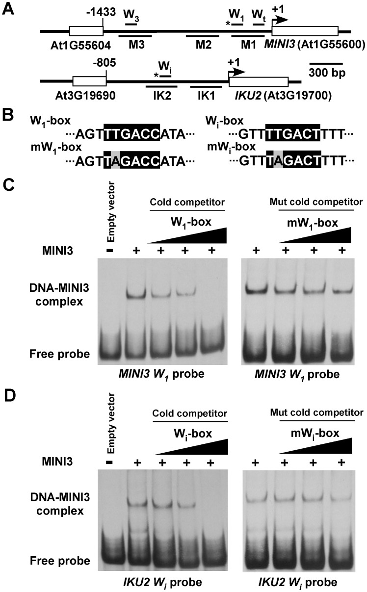
Figure 5. MINI3 binds to W-boxes in the MINI3 and IKU2 promoters.
(A) A schematic diagram of the MINI3 and IKU2 loci, the five amplicons (M1, M2, M3, IK1, and IK2) used for the ChIP-qPCR analysis, and the position of the W-boxes in the MINI3 and IKU2 promoters. Rectangles represent genes and numbers indicate genomic nucleotide sequence coordination. Arrowheads indicate transcription start sites, and the asterisk indicates W-boxes recognized by MINI3. (B) The nucleotide sequences of the W1-box, mutated W1-box (mW1-box), Wi-box, and the mutated Wi-box (mWi-box). Core sequences are shaded in black, and mutated nucleotides are shaded in gray. EMSA analysis of the binding of MINI3 to W1-box in the MINI3 promoter (C) or Wi-box in the IKU2 promoter (D). Cold wild type or mutated W1-box and Wi-box competitors were used at a molar excess of 5X, 10X, or 50X.

