
Figure 1. Identifying surface binding pockets.
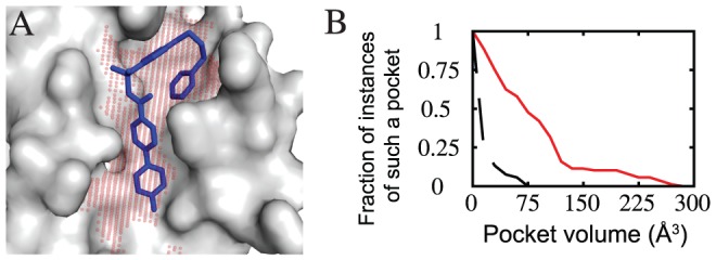
(A) Bcl-XL (grey surface) is shown in complex with an inhibitor (blue sticks). The protein surface features a large pocket (red spheres) complementary in shape to the inhibitor. (B) Deep pocket volumes of surface pockets at protein interaction sites harboring a bound inhibitor (red line) are larger than those found elsewhere on the protein surface (black line). Data are collected from a test set of seven proteins, each of which has been solved in complex with a small-molecule inhibitor (Bcl-XL, IL-2, FKBP12, HPV E2, ZipA, MDM2, and the BIR3 domain of XIAP).