
Figure 2. Surface pockets emerge only at druggable sites.
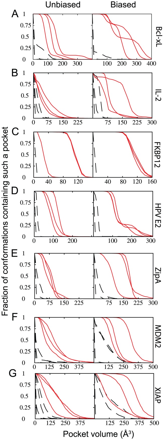
Volumes of surface pockets are shown from conformations generated with no biasing potential (left) and upon inclusion of a “pocket opening” biasing potential (right) for each of the seven proteins that comprise our test set. Surface pockets occur at druggable protein interaction sites (solid red lines) more frequently than elsewhere on the protein surface (dashed black lines). (A) Bcl-XL. (B) IL-2. (C) FKBP12. (D) HPV E2. (E) ZipA. (F) MDM2. (G) BIR3 domain of XIAP.