
Figure 5. The cell corpses of caspase-deleted mutants are cytologically and morphologically apoptotic.
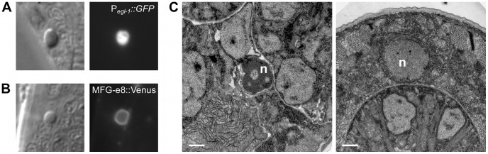
(A) Nomarski DIC and fluorescence images of a cell corpse within the head of a ced-1(e1735); csp-1(n4967); csp-2(n4871) ced-3(n3692) L1 larva carrying the integrated transgene nIs342[Pegl-1::gfp], a transcriptional reporter that expresses GFP under the control of the BH3 domain-only encoding gene egl-1. (B) Nomarski DIC and fluorescence images of a cell corpse within the head of a ced-1(e1735); csp-1(n4967); csp-2(n4871) ced-3(n3692) L1 larva carrying the extrachromosomal array nEx1646[Pdyn-1::mfg-e8::Venus], a fusion protein that binds to cell-surface exposed phosphatidylserine. (C) Representative transmission electron micrographs of cell corpses from ced-1(e1735); csp-1(n4967); csp-2(n4871) ced-3(n3692) larvae 24 hrs post hatching. “n”, nuclei of the cell corpses; scale bars, 0.5 microns. Note the difference in chromatin condensation between the two cell corpses.