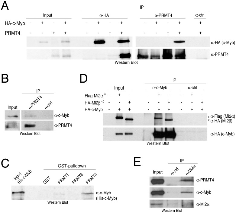
Figure 3. PRMT4 and Mi2 interact with the transcription factor c-Myb.
A: Co-immunoprecipitation of overexpressed PRMT4 with c-Myb. HEK293 cells were transfected with untagged PRMT4 and HA-c-Myb construct (alone or in combination) and harvested 48 hours after transfection. Protein extracts were subjected to IP using anti-HA (α-HA), anti-PRMT4 (α-PRMT4) antibodies or isotype control IgG (α-ctrl). Input (1%) and precipitates were stained by Western Blot analysis using anti-HA and anti-PRMT4 antibodies. B: Co-immunoprecipitation of endogenous PRMT4 with c-Myb. Jurkat cell extract was incubated with anti-PRMT4 (α-PRMT4) or isotype control IgG (α-ctrl). Input (5%) and precipitates were stained by Western Blot analysis using anti-c-Myb and anti-PRMT4 antibodies. The anti-c-Myb stainings for input and IP shown in the panels derive from the same blot and exposure time revealing that less than 5% of endogenous c-Myb interacts with endogenous PRMT4. C: PRMT4 and c-Myb are direct interaction partners. GST-pulldown with recombinant His-tagged c-Myb was performed using equal amounts of glutathione beads-bound recombinant GST, GST-PRMT1, -4 and -6 proteins. Input and bound His-c-Myb were visualised by Western Blot analysis using anti-c-Myb antibodies. D: Mi2α and Mi2β interact with c-Myb. HEK293 cells were transfected with HA-c-Myb plasmid together with Flag-Mi2α or HA-Mi2β. Protein extracts were incubated with anti-c-Myb antibodies or isotype control IgG (α-ctrl). Input and precipitates were stained by Western Blot analysis using anti-HA and anti-Flag antibodies. Asterisks indicate Flag-Mi2α and arrowheads indicate HA-Mi2β. E: Co-immunoprecipitation of endogenous Mi2α with PRMT4 and c-Myb. Jurkat cell extract was incubated with anti-Mi2α (α-Mi2α) or control IgG (α-ctrl). Input (5%) and precipitates were stained by Western Blot analysis using anti-PRMT4, anti-c-Myb and anti-Mi2α antibodies.

