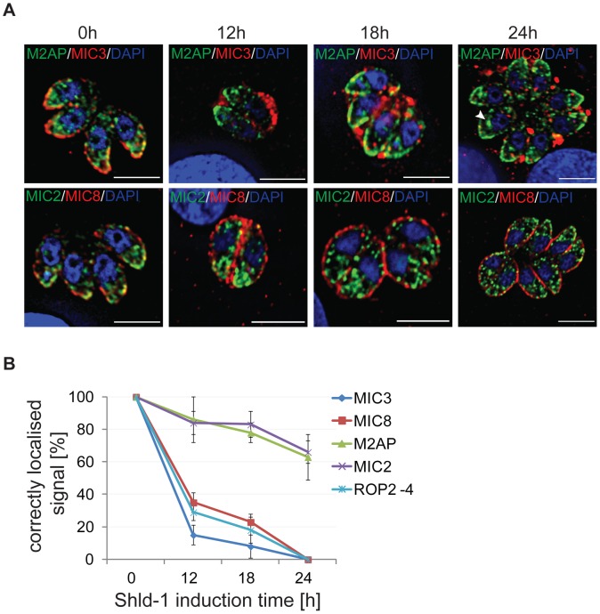
Figure 4. Time course analysis of parasites expressing ddFKBPmyc-Rab5A(N158I).
(A) Immunofluorescence analysis of intracellular parasites stably expressing ddFKBPmyc-Rab5A(N158I) treated for 0, 12, 18 and 24 hrs with 1 µM Shld-1 and co-stained with the indicated microneme antibodies (green/red) and Dapi (blue). (B) Quantification of localisations of Rop2-4 and indicated microneme proteins. 300–400 PVs of three independent experiments were analysed and normalised with RH hxgprt−parasites. Mean values and the respective standard deviation are presented. Note that MIC2 and M2AP is mainly mislocalised inside the parasite (arrowhead in A).

