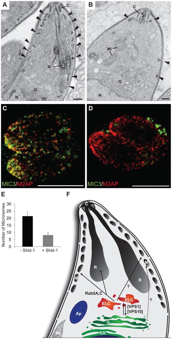
Figure 7. Ultrastructural and STED analysis of parasites expressing ddFKBPmyc-Rab5A(N158I).
(A) Electron microscopy analysis of the apical region of a tachyzoite expressing ddFKBPmyc-Rab5A(N158I) in the absence of 1 µM Shld-1 showing the natural morphological organisation of the apical organelles with various number of micronemes (arrowhead) associated with the limiting membrane complex. (B) Similar parasite stage from a culture incubated for 24 hrs with 1 µM Shld-1 showing a reduced number of micronemes (arrowheads). Scale bars represent 100 nm. (C,D) STED analysis of the same parasite strain grown in absence (C) or presence (D) of 1 µM Shld-1 for 18 hrs. Two-colour STED measurements were performed on MIC3 and M2AP, showing the absence of MIC3 within the parasites expressing ddFKBPmyc-Rab5A(N158I). (E) Electron microscopy quantification of micronemes present in longitudinal sections passing through the conoid and nucleus. 20 parasites per situation were quantified. Less micronemes are detected when ddFKBPmyc-Rab5A(N158I) is expressed. (F) Model of the vesicular traffic of secretory proteins to the rhoptries and micronemes. After modification at the Golgi secretory proteins are transported to the ELC's, which is regulated by VPS1 and VPS10. From the ELC's some of the microneme proteins (i.e. MIC3, MIC8, MIC11) and rhoptry proteins are transported to their target organelles in a Rab5A/C dependent manner. The regulation of the transport of other microneme proteins (AMA1, MIC2, M2AP, PLP1) remains unknown (?). VPS10 = Sortilin (TgSORTLR), VPS1 = dynamin related protein B (DrpB), Ap = Apicoplast, C = Conoid, ELC = endosome like compartments, ER = Endoplasmatic Reticulum, G = Golgi, Mi = Mitochondrium, M = Micronemes, R = Rhoptries.

