
Figure 2. Amino acid and DNA sequence of 2H10.
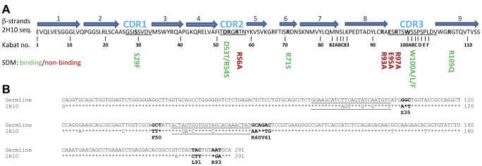
A) Amino acid sequence. Secondary structure assignment is based on the crystal structure. β-strands are depicted as blue arrows. CDR regions are indicated with CDR1 to CDR3 and are underlined. Residues depicted in bold were mutated. Mutants that still showed binding are shown in green and mutants that do not bind to the antigen anymore are shown in red. B) DNA sequence alignment of 2H10 with the germline V-gene from which it originated. The asterisks indicate identical nucleotides. The CDR1 and CDR2 coding regions are underlined. Codons with at least two point mutations are in boldface.