
Figure 7. Molecular modeling of the 2H10-gp41 peptide interaction suggests that 2H10 W100 is oriented towards the membrane.
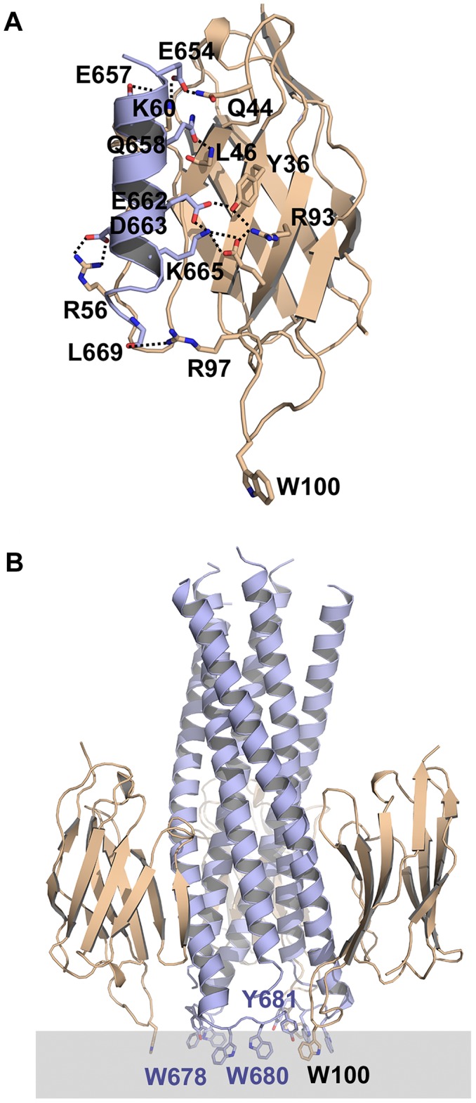
A) Molecular model of the gp41 peptide interaction with 2H10 produced by HADDOCK. Salt bridges and hydrogen bonds present in the top model and found in most of the top10 models are shown as dashed lines. B) Superpositioning of the Cα atoms of the gp41 peptide derived from the HADDOCK 2H10-peptide model onto the structure of a late fusion intermediate of gp41 [29] shows that the CDR3 W100 is oriented towards the membrane and found in the same plane as gp41 residues W678, W680 and Y681. Note that there are no clashes between the 2H10 VHH and gp41.