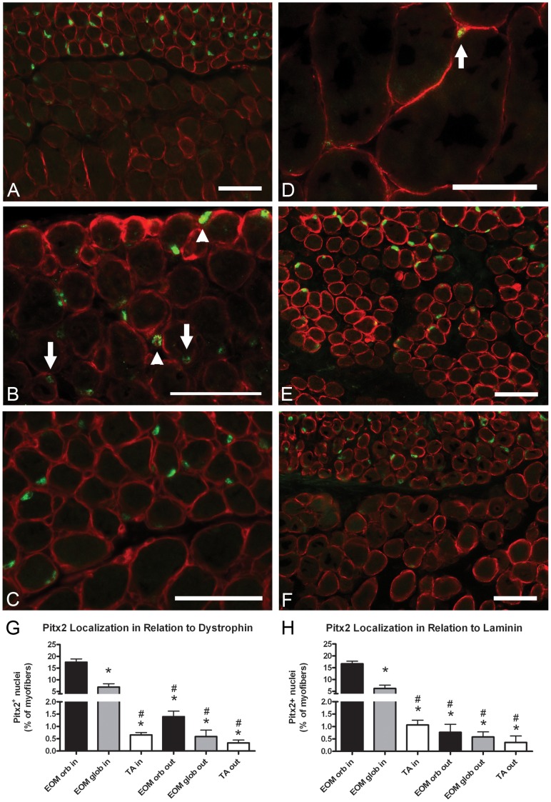
Figure 3. Pitx2-positive nuclei are located within myofibers and outside of myofibers in the satellite cell position.
(A, B) Pitx2 (green) and dystrophin (red) localization in wild-type mouse EOM. Arrows point down to Pitx2-positive myonuclei. Arrowheads point up to Pitx2-positive nuclei outside of the dystrophin ring. (C) Pitx2 (green) and laminin (red) localization in wild-type mouse EOM. (D) Pitx2 (green) and dystrophin (red) localization in wild-type mouse TA. Arrow points to a rare Pitx2-positive cell. (E) Pitx2 (green) and dystrophin (red) localization in human EOM. (F) Pitx2 (green) and dystrophin (red) localization in aged (19 months old) wild-type mouse EOM. (G) Percentages of Pitx2-positive nuclei in relation to dystrophin in wild-type mouse EOM and TA. (H) Percentages of Pitx2-positive nuclei in relation to laminin in wild-type mouse EOM and TA. orb, orbital layer of EOM. glob, global layer of EOM. Scale bars are 50 µm. * Indicates significant difference from EOM orbital inside (P≤0.05). # Indicates significant difference from EOM global inside (P≤0.05).

