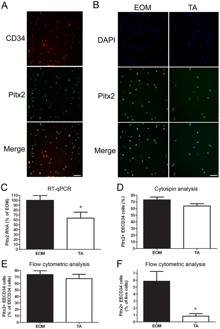
Figure 5. EECD34 cells express Pitx2.
(A, B) Pitx2 immunostaining of cytospun EECD34 cells from wild-type mouse EOM and TA. (C) Relative transcript levels of Pitx2 in cultured EECD34 cells from wild-type mouse EOM and TA. (D) Percentage of freshly isolated EECD34 cells from wild-type mouse EOM and TA that are Pitx2-positive determined by immunostaining of cytospun cells. (E) Percentage of freshly isolated EECD34 cells from wild-type mouse EOM and TA that are Pitx2-positive determined by flow cytometry. (F) Percentage of all freshly isolated mononuclear cells from wild-type mouse EOM and TA that are Pitx2-positive EECD34 cells determined by flow cytometry. Scale bars are 50 µm. * Indicates significant difference from EOM (P≤0.05).

