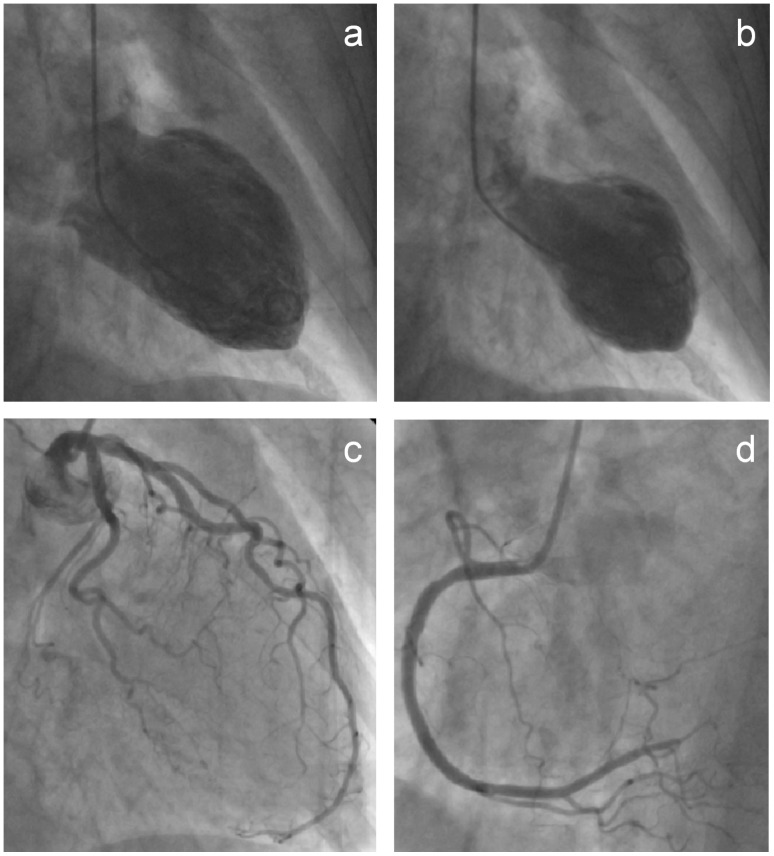
Figure 1. Typical left ventricular apical ballooning.
Left ventriculography in right anterior oblique projection during diastole (1a) and systole (1b) demonstrating akinesia of the apical and midventricular segments. Selective coronary angiograms of the left (1c) and right coronary artery (1d) excluding obstructive coronary artery disease.

