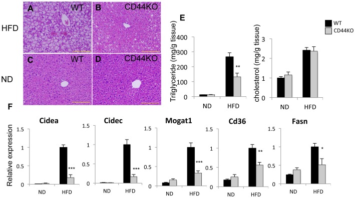
Figure 2. Hepatic steatosis was dramatically decreased in CD44KO(HFD).
A–D) Representative H&E stained sections of liver were presented. Scale bar indicates 200 µm. E) Comparison of hepatic triglyceride and cholesterol levels. F) Comparison of gene expression in the livers of WT or CD44KO mice fed a normal diet (ND) or a high fat diet (HFD) (n = 5–6 mice per each group). Data present mean±SEM, *p<0.05, **p<0.01, ***p<0.001.

