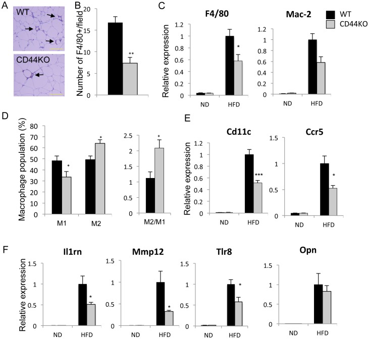
Figure 6. Adipose tissue inflammation was reduced in CD44KO(HFD) compared to WT(HFD) mice.
A) Macrophages in crown-like structures (CLS) were identified by immunohistochemical staining with an F4/80 antibody as indicated by arrows. Scale bar indicates 250 µm. B) The number of CLS was decreased in CD44KO(HFD) compared to WT(HFD) mice. F4/80 positive cells in at least 4 randomly selected fields in sections from 4 different mice were counted. C) Comparison of the expression of macrophage markers, F4/80 and Mac-2, in WAT of WT or CD44KO mice fed a normal diet (ND) or a high fat diet (HFD) (n = 5–6 mice per each group). D) SVF cells from WAT of WT(HFD) mice (n = 6) and CD44KO(HFD) mice (n = 8) were examined by FACS analysis with F4/80, CD11b, CD11c, and CD206 antibodies. The percentage and ratio of pro-inflammatory macrophage (M1: F4/80+CD11C+CD206−) and anti-inflammatory macrophage (M2: F4/80+CD11C−CD206+) were determined. Expression of several macrophage markers (E) and inflammatory markers (F) was analyzed in WAT of WT or CD44KO mice fed a normal diet (ND) or a high fat diet (HFD) (n = 5–6 mice per each group).

