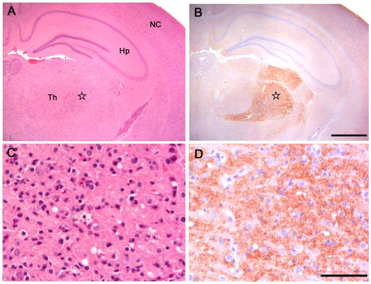
Figure 1. Histopathological and immunohistochemical analysis of Bv109I inoculated with Bv109ICWD.
(A) Serial sections showing neocortex (NC), hippocampus (Hp) and thalamus (Th), stained by haematoxylin and eosin and (B) by immunohistochemistry for PrPSc with SAF84 mAb - bar 500 µm. (C) and (D): magnification of the ventral thalamic nucleus (star) from (A) and (B), respectively - bar 50 µm.

