
Figure 2. Lesion profiles of Bv109I infected with CWD following primary transmission and third passage.
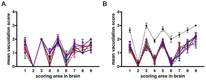
Pathological phenotypes observed after primary transmission and third passage are shown in (A) and (B), respectively. CWD1, 2 and 4 from elk, are shown in red, green and violet lines with open circles, respectively; CWD3 from mule deer, in blue line and closed circles; CWD5, 6 and 7 from W-T deer in red, green and violet dashed lines and closed triangles, respectively. Vole-adapted sheep scrapie is shown in black dashed line and closed circles. Brain-scoring areas are: medulla (1), cerebellum (2), superior colliculus (3), hypothalamus (4), thalamus (5), hippocampus (6), septum (7), retrosplenial and adjacent motor cortex (8), cingulate and adjacent motor cortex (9).