
Figure 3. Western blot analysis of the PrPres level in the brain of CWD-affected voles.
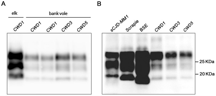
(A) Immunoblot of proteinase K–resistant PrPSc (PrPres) from one original elk isolate (CWD1) and from voles infected with CWD1, 3 and 5. (B) Comparison of PrPres amount and glycoprofile between Bv109I-adapted CWD and Bv109M-adapted sporadic CJD, sheep scrapie and cattle BSE, at third passage. Membranes were probed with SAF84. Molecular size markers are shown in kilodaltons on the right.