
Figure 4. Regional distribution, by PET-blot, of PrPres following the third passage of CWD1, CWD3, CWD5 and sheep scrapie in Bv109I.
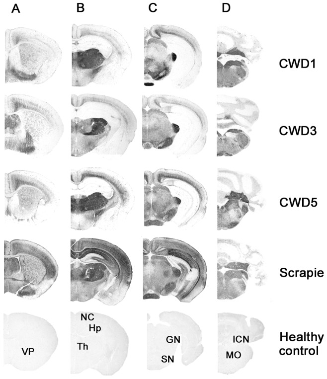
Coronal sections of the forebrain representing: telencephalon (A), diencephalon (B), midbrain (C) and hindbrain (D). In the lower part of the figure, the labelled coronal sections of negative control brain from 150 days old Bv109I are shown: VP, ventral pallidum; NC, neocortex; Hp, hippocampus; Th, thalamus; GN, geniculate nuclei; SN, substantia nigra; ICN, interposed cerebellar nucleus; MO, medulla oblongata.