
Figure 6. Differentiation of hESC to NPCs and mature neurons.
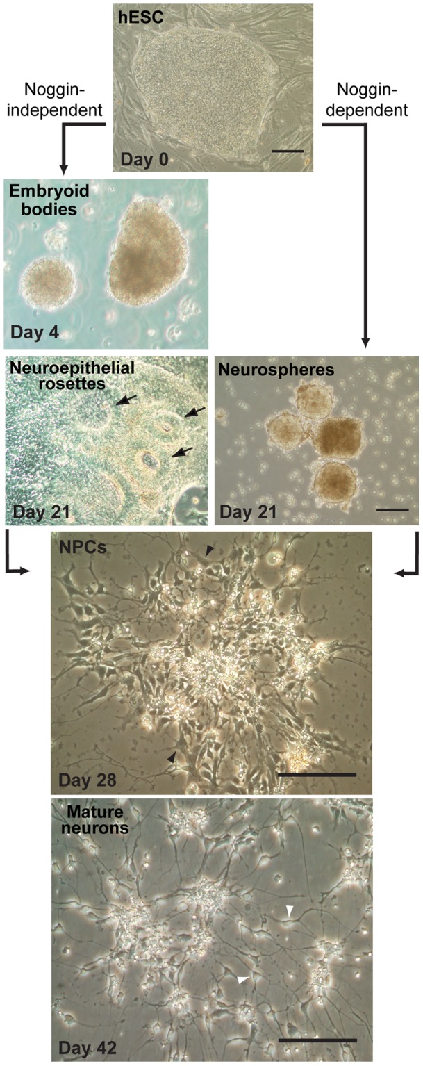
Phase-contrast micrographs of a pluripotent hESC colony growing on a layer of irradiated mouse embryonic fibroblasts on day 0 (top image), subsequent differentiation in the absence of noggin into embryoid bodies on day 4 and neuroepithelial rosettes on day 21 (left images) or in the presence of noggin into neurospheres in suspension culture on day 21 (right image), and final differentiation into adherent cultures enriched in NPCs on day 28 and mature neurons by day 42 (bottom images). Arrows in day 21 noggin-independent cultures indicate neuroepithelial rosettes. Open arrowheads in day 28 NPC cultures indicate characteristic cells with large perikaryon and minimal processes, whereas closed arrowhead in day 42 mature neuron cultures indicate characteristic cells with small perikaryon and extensive processes. Scale bars = 200 μm.