
Figure 3. 46-year-old woman with multiple thyroid nodules.
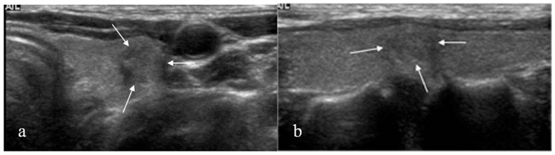
US (a: transverse, b: longitudinal) revealed a 8-mm sized hypoechoic nodule (arrows) with peripheral calcifications in the mid pole of left thyroid. US assessment was suspicious malignant. Initial US-FNA showed non-diagnostic cytology and negative for BRAFV600E mutation. This nodule was diagnosed as suspicious for malignancy on follow-up US-FNA performed 11 months later, and positive for BRAFV600E mutation. Surgery confirmed this lesion as PTC.