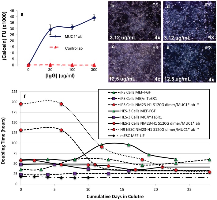
Figure 4. Anti-MUC1* antibodies are a novel surface coating that enables stem cell attachment and growth.
a) An anti-MUC1* rabbit polyclonal antibody or a control IgG antibody were adsorbed at varying concentrations onto a tissue culture treated multi-well plate. BGO1V/hOG hES cells were plated onto the surfaces and allowed to grow for 48 hours. A Calcein AM assay to quantify cell number was performed. Anti-MUC1* antibody, but not the control antibody, enabled stem cell attachment and growth. BGO1V/hOG hES cells were cultured for 20 passages in NM23-H1-MM without a decrease in pluripotency or change in karyotype (data not shown). b–e) H9 hES cells (b, c) or iPS cells (d, e) were plated onto VitaTM multi-well plates, reported to have higher protein binding capability, that were coated with either 3.12 µg/mL (b, d) or 12.5 µg/mL (c, e) of a monoclonal anti-MUC1* antibody, MN-C3. Cells attached and proliferated as a function of antibody concentration, with maximal attachment observed at 12.5 µg/mL. f) Doubling times forhuman ES cells (H9 or HES-3), human iPS cells and mouse ES cells were measured as a function of various culture media and surfaces. The traces marked by red circles/dotted line and red circles/dashed line (human H9 ES and iPS cells, respectively) show the change in doubling time as cells transition from bFGF-based media to NM23-H1 media on anti-MUC1* surface.

