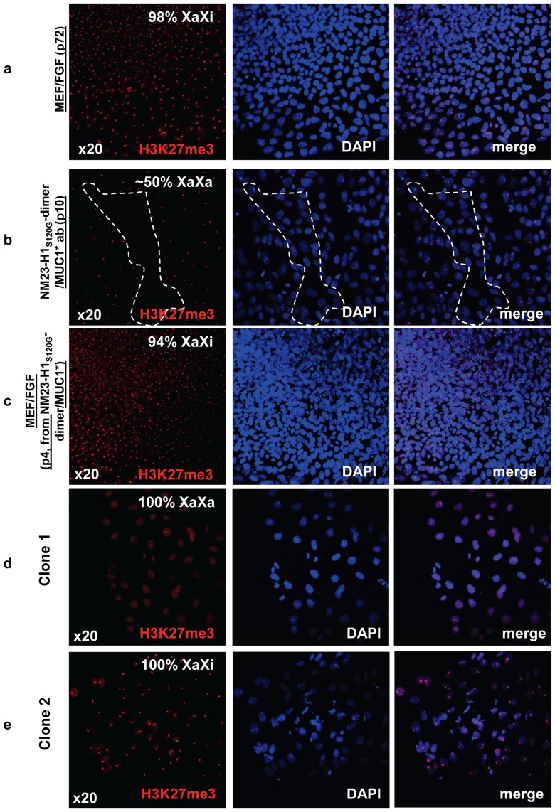
Figure 7. hES cells cultured in NM23-H1-MM on Anti-MUC1* surfaces are pre X-inactivation, characteristic of naïve state.
a) Staining with nuclear marker DAPI and antibodies against tri-methylated (Lys27) Histone 3 shows that ES cells cultured in bFGF over MEFs are XaXi or post X-inactivation, characteristic of primed state. b) The cells shown in (a) were then transferred to anti-MUC1* antibody surfaces and cultured in NM23-H1 media for 10 passages then stained for X-activation status as in (a). The confocal images show a 50/50 mix of regions that are XaXa (dotted region) and others that are XaXi. c) The ES cells shown in (b) were then transferred back again to culture in bFGF over MEFs for 4 passages and images show 95% reversion to the XaXi, characteristic of primed state. d, e) ES cells cultured in NM23-H1 media over an anti-MUC1* antibody surface for 14 passages were serially diluted and allowed to grow until isolated colonies were observed. Cells were stained with nuclear marker DAPI and antibodies against tri-methylated (Lys27) Histone 3 to measure Chromosome-X status. X-activation status was clonal as we isolated clones with 100% X-inactivated (XaXi) and clones with 100% X-activated (XaXa).

