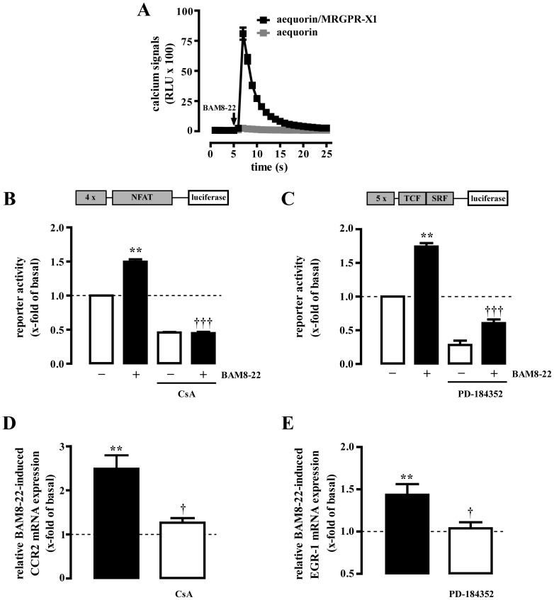
Figure 6. MRGPR-X1 induce EGR-1 via ERK-1/2 and CCR2 via NFAT in primary DRG neurons.
BAM8-22-induced (2 µM) calcium signals in rat DRG neurons co-expressing MRGPR-X1 and aequorin or solely aequorin are presented in (A). BAM8-22-induced (2 µM, 8 h) activation of the NFAT (B) or TCF/SRF (C) reporter is shown in rat DRG neurons transiently co-expressing MRGPR-X1. RTQ-PCR experiments were performed with cDNAs derived from serum-starved MRGPR-X1 expressing rat DRG neurons stimulated or not with BAM8-22 (2 µM) for 6 h (D) or 40 min (E). CsA (1 µM, 30 min) was used to block calcineurin in (B and D) or PD-184352 (10 µM, 30 min) to inhibit ERK-1/2 activity in (C and E). Relative BAM8-22-induced gene expression was normalized to β-actin and calculated using the ΔΔCp method. Data from 4 independent experiments were compiled and expressed as the mean ± S.E.M. Asterisks indicate a significant difference to not stimulated cells. Dagger signs indicate a significant difference between BAM8-22-stimulated inhibitor-treated and untreated cells.

