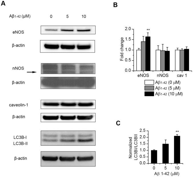
Figure 6. Affects of exogenous Aβ1–42 on the level of eNOS and autophagy in endothelial cells.
Endothelial cells were treated with 5 µM and 10 µM Aβ1–42 and incubated for 12 hrs. Protein levels of eNOS, nNOS, caveolin-1 and the autophagy marker LC3B were analyzed by Western blotting (A). (B), quantitation of protein levels of eNOS, nNOS and caveolin-1 normalized to β-actin control. Results are mean ± SD (n = 3–5). (C), quantitation of LC3B II/I ratio normalized to β-actin control. Results are mean ± SD (n = 3). **, P<0.01 compared to control.

