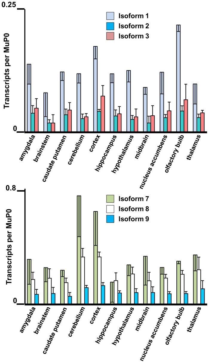
Figure 7. Highly expressed Fmr1 transcript isoforms in eleven brain regions.
The amygdala, brainstem, caudate putamen, cerebellum, cortex, hippocampus, hypothalamus, midbrain, nucleus accumbens, olfactory bulb and thalamus were dissected from three adult mouse brains. The levels of the abundant Fmr1 transcripts are shown with standard deviations in biological triplicates.

