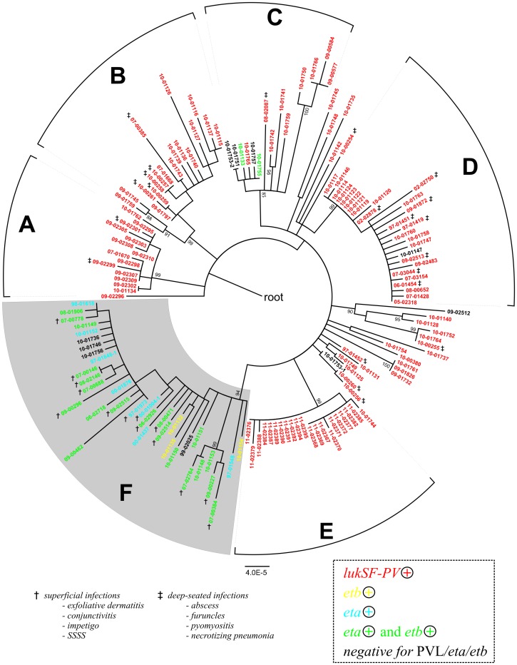
Figure 3. Toxin gene complement and clinical phenotype.
Maximum likelihood phylogenetic tree as in Figure 1, indicating the presence of toxin genes lukSF-PV, eta, and etb (colours) and clinical phenotype where this was reported (symbols: †, superficial infection, i. e., impetigo, staphylococcal scalded skin syndrome, conjunctivitis, or exfoliative dermatitis); ‡, deep-seated infection (abscess, furuncle, pyomyositis, necrotizing pneumonia).

