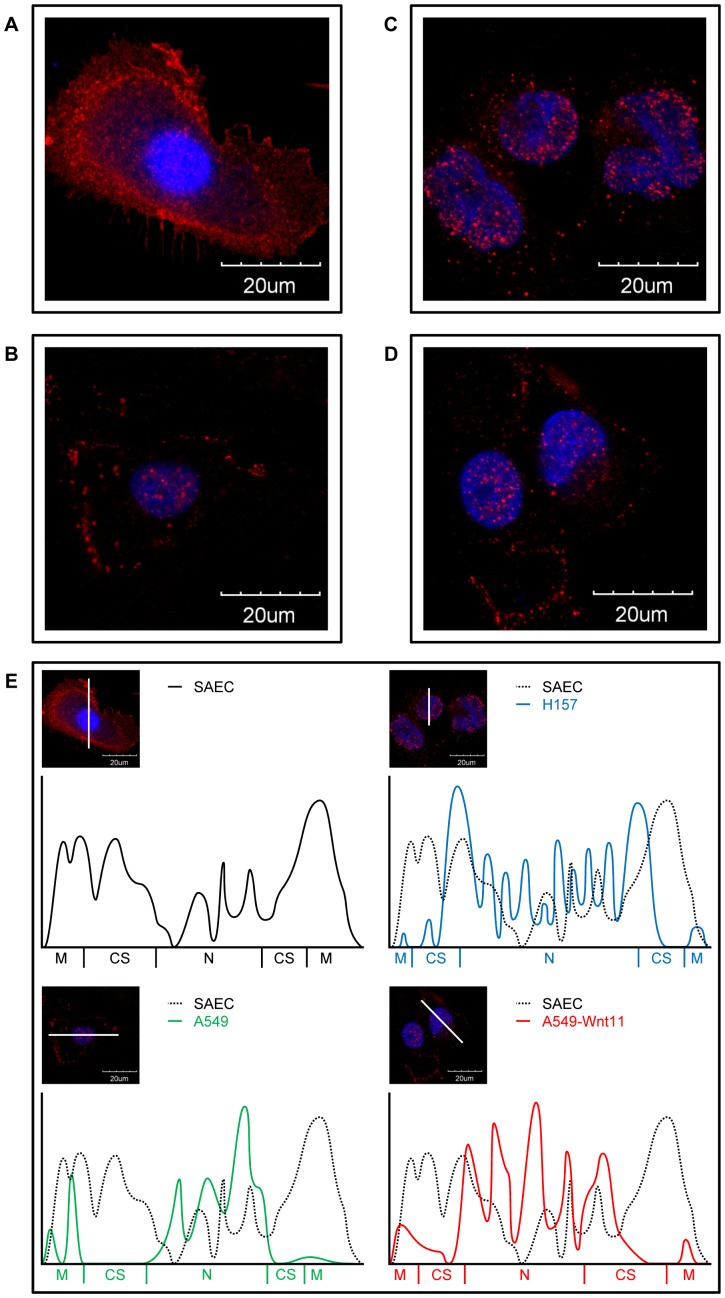
Figure 4. Localization of β-catenin. A:
Immunofluorescent staining of SAEC. B: Immunofluorescent staining of normal A549. C: Immunofluorescent staining of Wnt11 overexpressing A549. D: Immunofluorescent staining of H157 monolayer cell cultures. (60x image, red: β-catenin, blue: DAPI). Note the dramatic increase in nuclear localization and the decrease in cellular membrane localization of A549 AC, Wnt11-A549 and H157 SCC cell lines compared to the normal pulmonary epithelium (SAEC). Data presented are representative of three independent experiments. E: Densitometry of immunofluorescent images of SAEC, A549, Wnt-11-A549 and H157 cells. Note the increased nuclear localization of β-catenin particularly in the Wnt11-A549 cell line. (M: cellular membrane, CS: cytosol, N: nucleus).

