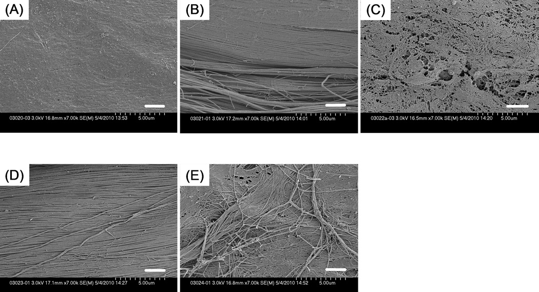
Figure 7.
SEM images at higher magnification (× 7000) showing tendon surface that underwent each surface treatment. (A) untreated tendon; (B) tendon with mechanical abrasion; (C) tendon with trypsin treatment; (D) tendon with combined treatment; (E) untreated extrasynovial tendon. The mesh-like structure of epitenon layer is exposed in trypsinized tendon. Scale Bar: 2 µm.

