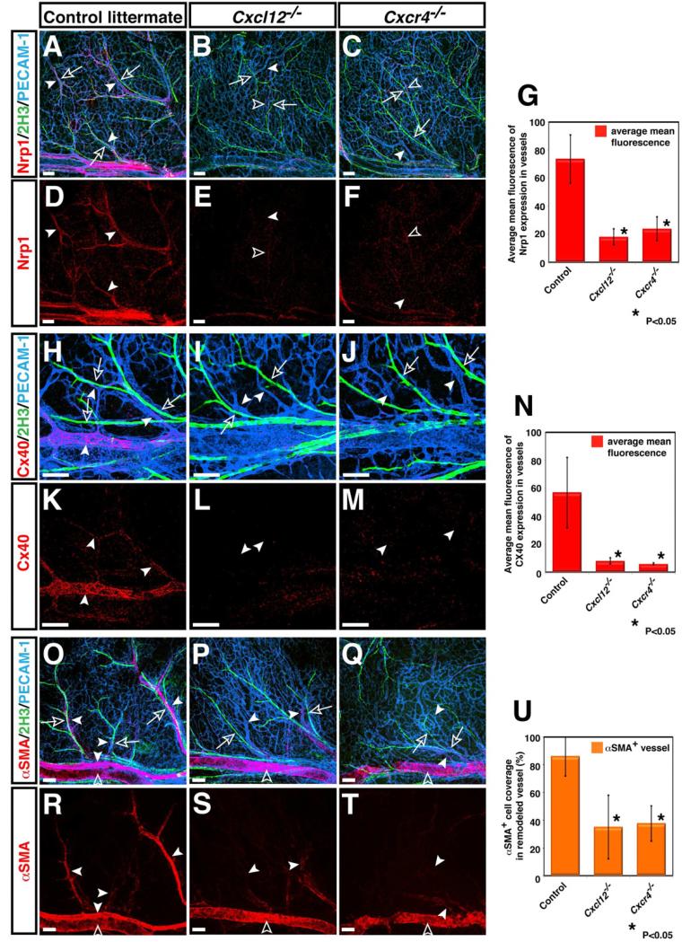
Figure 3. Defective arterial differentiation and smooth muscle coverage in Cxcl12 and Cxcr4 homozygous mutants.
Whole-mount triple immunofluorescence labeling of limb skin with antibodies to arterial markers either Nrp1 (A-F, red, arrowheads) or Cx40 (H-M, red, arrowheads) and the smooth muscle cell marker αSMA (O-T, red, arrowheads), in addition to PECAM-1 (A-C, H-J and O-Q, blue) and neurofilament (2H3, A-C, H-J and O-Q, green, open arrows) in Cxcl12-/- (B, E, I, L, P, S), Cxcr4-/- (C, F, J, M, Q, T) or control littermates (A, D, H, K, O, R) at E15.5 is shown. The expression of arterial markers such as Nrp1 and Cx40 was conspicuously reduced in remodeled vessels of both Cxcl12-/- and Cxcr4-/- mutants (B, C, E, F for Nrp1 expression; I, J, L, M for Cx40 expression, arrowheads). Note that weak Nrp1 expression can be detected in some remodeled vessels in relatively close proximity to nerves in Cxcl12-/- (B, E, open arrowheads) and Cxcr4-/- mutants (C, F, open arrowheads). Fewer αSMA+ smooth muscle cells associated with small-diameter branched vessels in Cxcl12-/- and Cxcr4-/- mutants (O versus P and Q, R versus S and T, arrowheads), although smooth muscle coverage in large-diameter veins appeared unaffected (S and T, open arrowheads). Quantification of arterial marker expression (G and N) or αSMA+ smooth muscle cell coverage (U) in small-diameter branched vessels was performed (n=4 per genotype; bars represent mean ± SEM). Asterisk indicates statistically significant difference (P<0.05) in both mutants compared with control littermates according to Student's t-test. Scale bars are 100 μm.

