FIGURE 7.
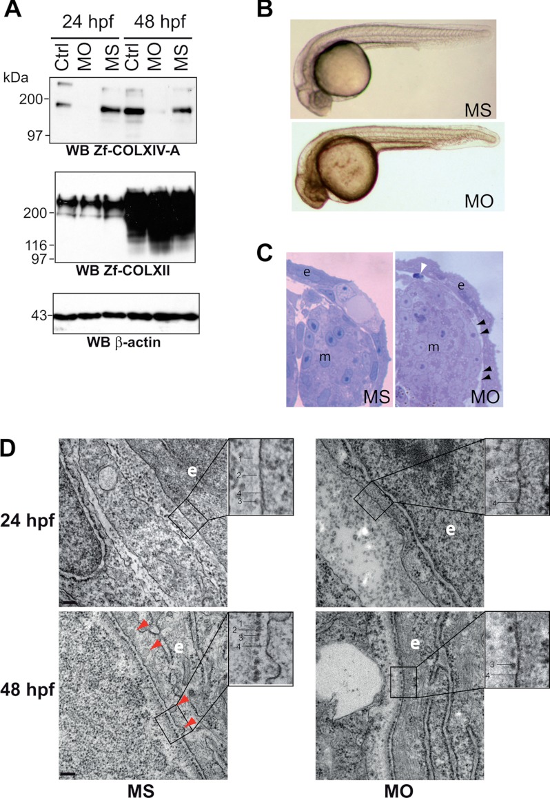
COLXIV-A down-regulation in zebrafish embryos leads to defects in basement membrane formation. A, validation of morpholino-induced COLXIV-A down-regulation. AB/Tu eggs were injected at one cell stage with 4.2 ng of control MS or MO, and collagen expression was analyzed by Western blot. COLXIV-A protein expression was significantly decreased at 24 and 48 hpf in embryos injected with morpholinos against col14a1a (MO) as compared with MS-injected embryos whereas COLXII was not affected (n = 3). B, knockdown of COLXIV-A expression had no effect on gross morphology of embryos. Injected embryos were analyzed by stereomicroscopy at 24 hpf (n = 5 separate experiments with at least 100 injected embryos/condition in each experiment). C, knockdown of COLXIV-A expression caused skin detachment. Transversal thick sections of 24-hpf MO- and MS-injected embryos were stained and analyzed by stereomicroscopy. At this stage, compared with the MS-injected embryos, MO-injected embryos showed zones of epidermal detachment (e), resulting in gaps between epidermis and adjacent tissue (black arrowheads). In addition, apoptotic cells (white arrowhead) were visible beneath the skin. m, muscle. The pictures correspond to the right upper part of the embryos at the yolk sac extension level. D, knockdown of COLXIV-A expression impairs basement membrane formation. Transmission electron microscope images of MS- and MO-injected embryos at 24 and 48 hpf. At 24 hpf, a basement membrane consisting of electron dense (lamina densa, line 1) and electron lucent (lamina lucida, line 2) material can be discerned in MS-injected embryos. In addition, sparse adepidermal granules (line 3) are visible in the lamina lucida of MS-injected embryos. In contrast, the lamina lucida and lamina densa are missing in MO-injected embryos at 24 hpf, although some adepidermal granules (line 3) can be discerned close to the basal plasma membrane (line 4). At 48 hpf, adepidermal granules (line 3) are homogenously deposited in the lamina lucida (line 2) of MS-injected embryos and thus form a nice sheet-like structure. In contrast, in MO-injected embryos, the adepidermal granules are irregularly spaced, showing frequent gaps. Furthermore, the lamina lucida is very thin, and the lamina densa has a fuzzy appearance, indicating defects in BM structure. Moreover, invaginations of the epithelial cell plasma membrane (line 4) observed in the MS-injected embryos are less frequent in the morphants (red arrowheads). Scale bars: 24 hpf, 100 nm; 48 hpf, 200 nm. WB, Western blot; Ctrl, control.

