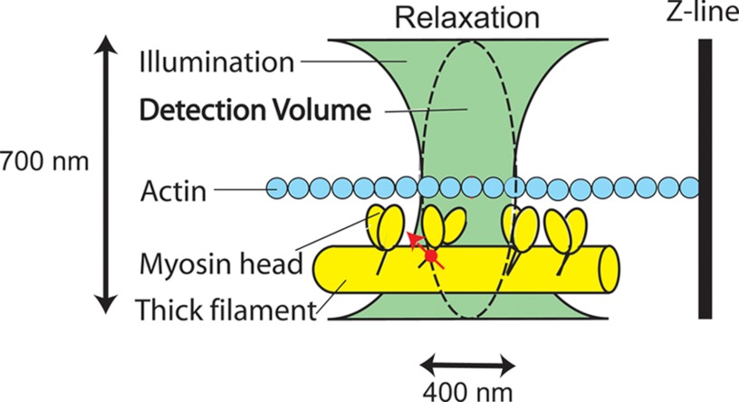
FIGURE 2.
Testing the SRX hypothesis. Myosin light chain 1 is labeled with fluorescent dye (red). A small volume of a myofibril is illuminated through a high numerical aperture objective (with the green light) to excite the fluorophore. The detector sees only the confocal volume (∼1 μm3) equivalent to the ellipsoid of revolution indicated by the dashed line (see ”Materials and Methods“).

