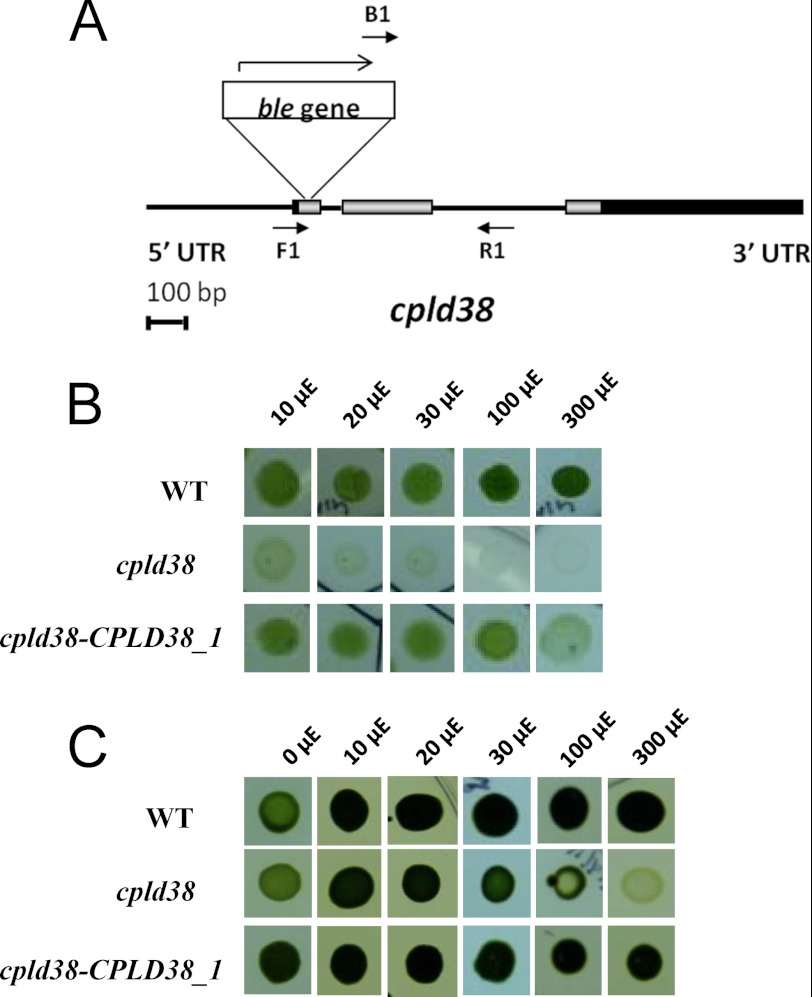
FIGURE 2.
cpld38 mutant and its responses to light under photoautotrophic and mixotrophic conditions. A, map showing insertion of the ble gene in the cpld38 mutant. Exons are shown as gray boxes, introns and intergenic regions as black lines, and the 5′- and 3′-UTRs as black boxes. The ble gene is inserted into the first exon, as indicated. Primers used for the analysis of progeny from genetic crosses are represented by short arrows (see supplemental Fig. S3). B and C, WT cells, cpld38, and the cpld38-CPLD38_1 rescued mutant were grown to exponential phase in TAP medium and then diluted to 1 × 10⁵ cells ml−1. Five μl of culture was spotted onto solid (1.5% agar), minimal (B), or TAP (C) medium. Cultures were grown at the light intensities indicated for either 10 days (minimal medium) or 7 days (TAP medium). The designation μE is equivalent to μmol photons m−2 s−1.

