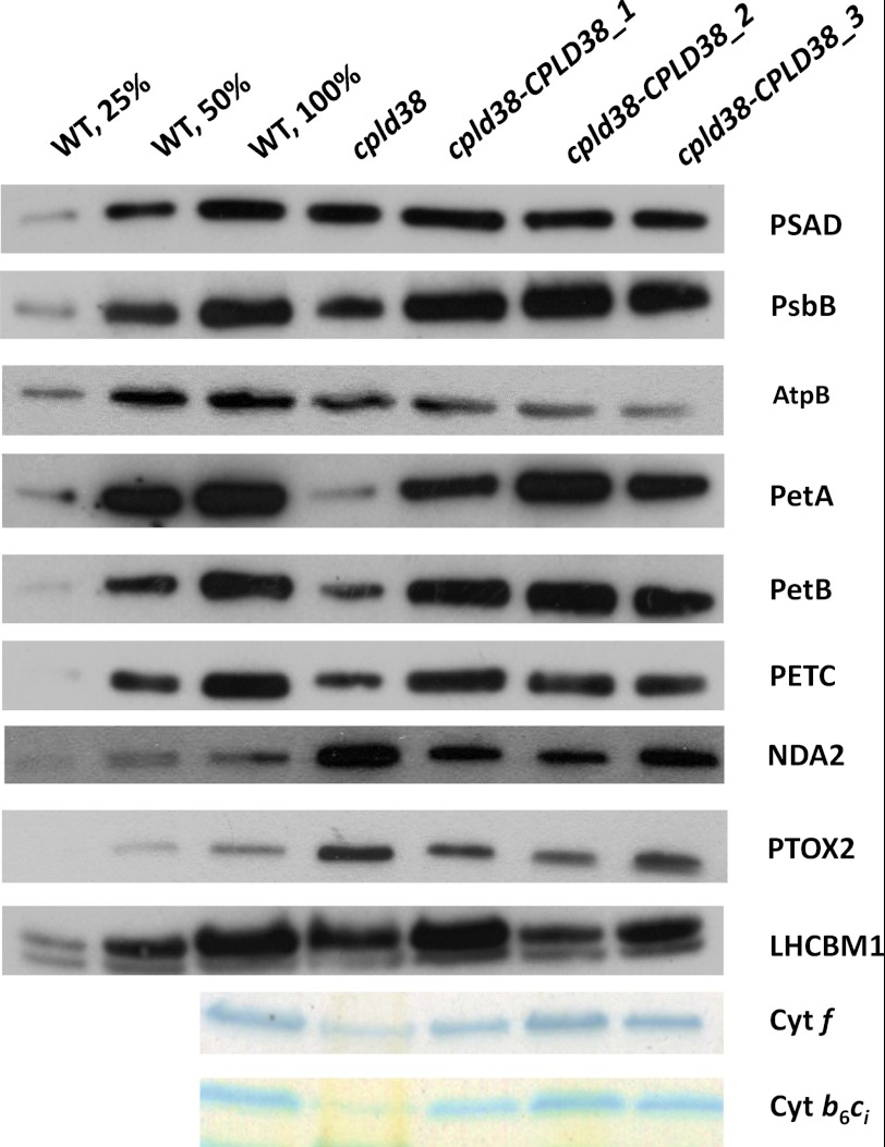
FIGURE 5.
Western blot and heme staining analyses to identify photosynthetic and chlororespiratory components. Chlamydomonas proteins from the WT, cpld38, and the various cpld38-CPLD38 rescued strains were resolved by SDS-PAGE on a 15% polyacrylamide gel and detected immunologically or by heme staining. Samples were taken for analysis when the strains were in their exponential growth phase in low light (40 μmol photons m−2 s−1) on TAP media with moderate shaking (150 rpm). The antibodies used for this analysis are against polypeptides indicated on the right side of the figure. Approximately 1 μg of chlorophyll was loaded for each sample analyzed by Western blotting, whereas∼30 μg of chlorophyll was loaded for each sample analyzed for heme-containing polypeptides. The LHCBM1 protein was used as a loading control.

