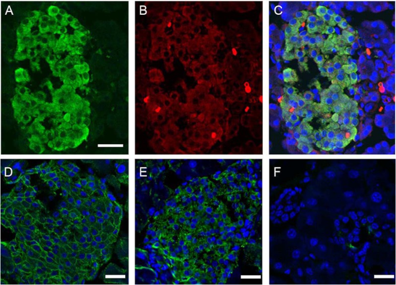
FIGURE 3.
Immunocytochemical localization of FXYD2 in rodent pancreas. Representative confocal images of rat pancreatic islet double-labeled for insulin (A) and FXYD2b (B) are shown. Secondary antibodies were FITC-conjugated anti-guinea pig (A) and Alexa-555 anti-rabbit (B) antibodies. Co-localization is seen in the merged image (C). Counterstaining for nuclei (blue, C) was with To-Pro. Bar, 20 μm. D–F, shown is immunostaining of mouse pancreatic islet with polyclonal rabbit antibodies against α1 subunit of Na,K-ATPase (green, D) and FXYD2b (green, E). Secondary antibodies were donkey-anti-rabbit Alexa 488. F, shown is the negative control for secondary antibodies alone. Nuclei (D–F) were counterstained with To-Pro. Bars, 20 μm.

