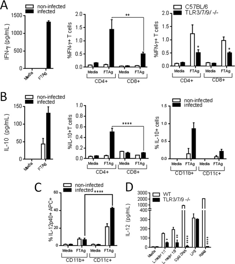
FIGURE 6.
Characterization of early cellular immune response upon infection with L. major. Wild type C57BL/6 mice were infected in the right footpad with 106 metacyclic forms of the L. major and, at 12 days after inoculation, animals were sacrificed, and the draining lymph nodes of the infected footpad were obtained. Cells were grown in absence (medium) or presence of a total lysate of L. major metacyclic parasites (FTAg) 48 h, and supernatant was used for quantification of IFN-γ (A), IL-10 (B), and IL-12 (C) by ELISA. Analysis by flow cytometry of CD4+ and CD8+ cells from WT and TLR3/7/9−/− mice 12 days after infection is also shown. B and C, T cells expressing CD4+ and CD8+ surface markers were stained for endogenous IFN-γ and IL-10, and cellular populations were separated by flow cytometry, as well CD11b+ and CD11c+ cells that were tested for IL-10 and IL-12 (please refer to “Experimental Procedures” for all cytokines tested). E, data represent the mean ± S.E. (n = 3). Differences are considered statistically significant (*, p < 0.05; **, p < 0.01; and ****, p < 0.0001) after two-way ANOVA with Bonferroni's post-test, are indicated.

