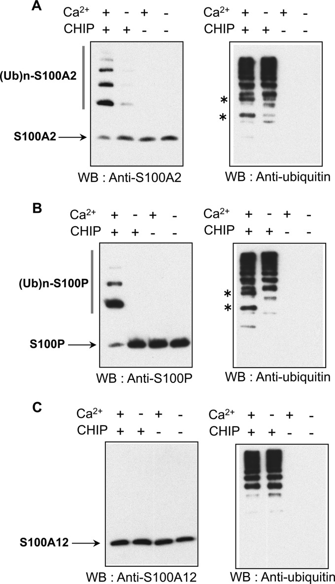
FIGURE 6.
S100 protein itself is ubiquitinated by CHIP in a Ca2+-dependent manner. A–C, purified 10 μm S100A2 (A), S100P (B), and S100A12 (C) were subjected to in vitro ubiquitination assays with (+) or without (−) 1 mm CaCl2 (Ca2+) and CHIP. The details are described under “Experimental Procedures.” The samples were analyzed by Western blotting (WB) with anti-specific S100 antibodies (left panel) and anti-ubiquitin antibody (right panel) as indicated. Asterisks indicate significant ubiquitin conjugates of S100 proteins. Unmodified S100A2, S100P, and S100A12 are indicated by arrows. Ubiquitylated S100 proteins are shown as (Ub)n-S100A2 and (Ub)n-S100P. The data shown in each panel are representative of three independent experiments. (Ub)n-S100A2, ubiquitylated S100A2; (Ub)n-S100P, ubiquitylated S100P.

