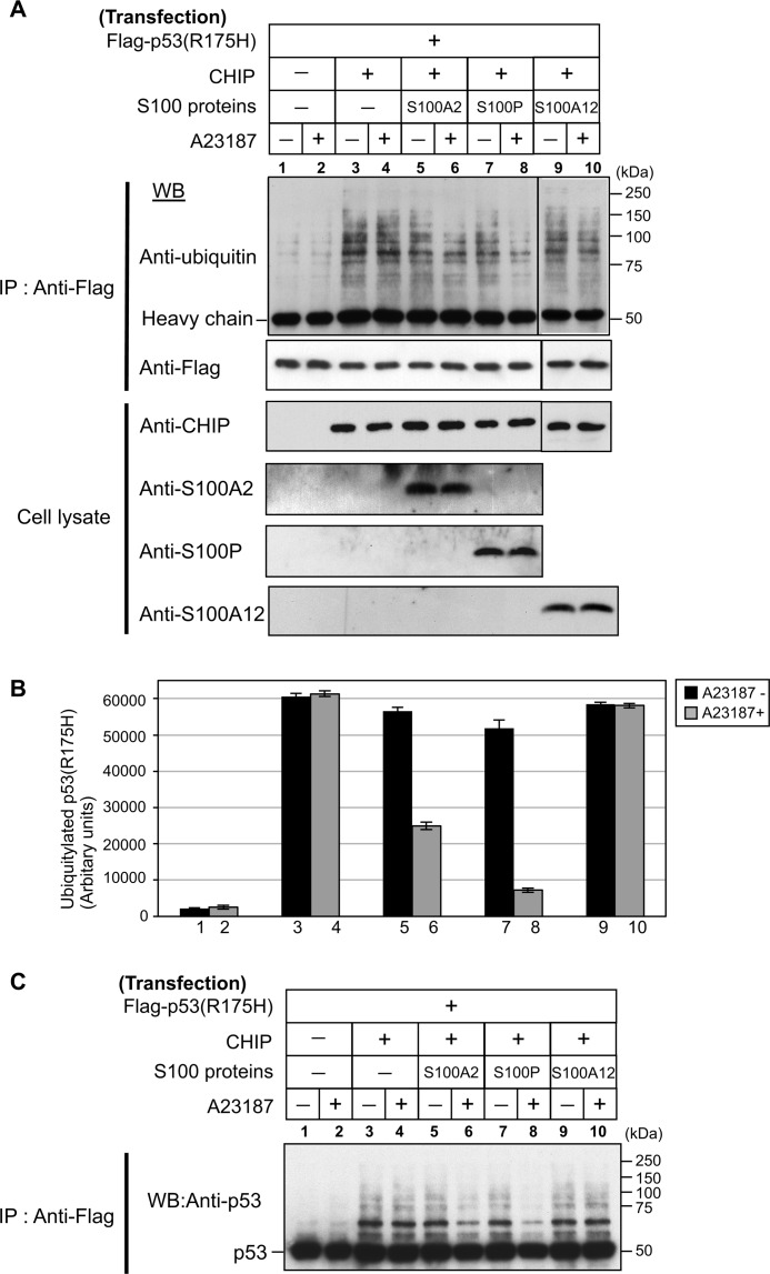
FIGURE 7.
Ca2+/S100 proteins suppress the CHIP-mediated ubiquitination of mutant p53 in vivo. A, Hep3B cells were transiently transfected with (+) or without (−) FLAG-p53R175H, CHIP, S100A2, S100P, and S100A12. The transfected components of each dish are indicated on the top panels. Transfected Hep3B cells were treated with (+) or without (−) 1 μm A23187 for 6 h. Cell lysates were immunoprecipitated (IP) with anti-FLAG antibody. Details are described under “Experimental Procedures.” Lysates and immunoprecipitated proteins were detected by Western blotting (WB) with the indicated antibodies. Molecular mass markers are indicated on the right. B, the ubiquitination levels of p53R175H with A23187 (gray) or without A23187 (black) are plotted. The error bars represent the S.E. with n = 3. C, immunoprecipitated proteins were detected by Western blotting (WB) with anti-p53 antibody. Molecular mass markers are indicated on the right.

