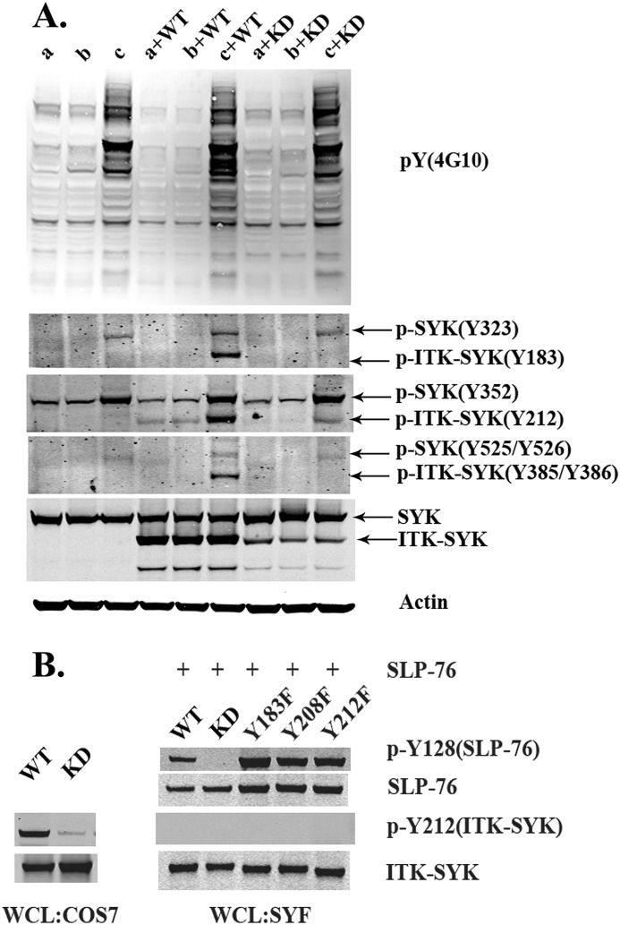
FIGURE 6.
Phosphorylation of ITK-SYK in T lymphocytes and in SYF cells. A, Western blot analysis of Jurkat cells transfected with ITK-SYK or ITK-SYK-KD. Following transfection, cells were processed for immunoblotting and decorated with phosphospecific antibodies as described in Fig. 1B. Cells were either serum-starved (2 h) or treated with pervanadate (100 μm, 20 min on ice). Minus serum (lane a), steady state (lane b), and pervanadate (lane c). The upper panel shows global tyrosine phosphorylation, and the lower panel shows phosphorylation of critical tyrosines in endogenous SYK, ITK-SYK, and kinase-inactive ITK-SYK. B, Western blot analysis of SYF cells, lacking SFKs, transfected with ITK-SYK and ITK-SYK mutants. (For details regarding constructs, see Table 1.) Lysates from COS7 cells transfected with ITK-SYK or ITK-SYK-KD were run in parallel as control for detection of ITK-SYK phosphorylation at Tyr-212.

