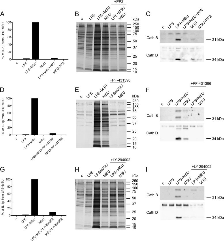
Fig. 5.
MSU-induced protein secretion is dependent on the activity of Src, Pyk2, and PI3 kinases. Macrophages were left untreated or primed with LPS after which they were activated with MSU for 3 h in the absence and presence of Src kinase inhibitor PP2 (10 μm). After this the cell culture supernatants were collected. A, The secretion of IL-1β was measured by ELISA. The values obtained in LPS and MSU co-stimulated cells were set at 100%. Representative data from one experiment is shown; n = 3 independent experiments. B, Secreted proteins were visualized with silver staining and (C) the secretion of Cathepsins B and D was analyzed by Western blotting. Macrophages were left untreated or primed with LPS after which they were activated with MSU for 3 h in the absence and presence of Pyk2 kinase inhibitor PF-431396 (10 μm). D, The secretion of IL-1β was measured by ELISA. Representative data from one experiment is shown; n = 2 independent experiments. E, Secreted proteins were visualized with silver staining and (F) the secretion of Cathepsins B and D was analyzed by Western blotting. Macrophages were activated with LPS and MSU as described with or without PI3K inhibitor LY-294002 (100 μm). G, The secretion of IL-1β was measured by ELISA. Representative data from one experiment is shown; n = 2 independent experiments. H, Secreted proteins were visualized with silver staining, and (I) the secretion of cathepsins was analyzed by Western blotting.

