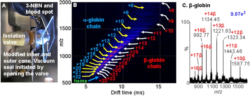
Fig. 4.
MAIV-IMS-MS of a solvent-extracted blood spot from a Band-Aid using a commercial atmospheric pressure ESI source with a modified skimmer cone to provide a larger inlet aperture. A, photograph of the device showing the isolation valve in the open position with a glass microscope slide held by the pressure differential against the skimmer opening, with the matrix–blood spot sample exposed to the vacuum initiating ionization; more details on source modifications are provided in supplemental Fig. S1. B, two-dimensional IMS-MS plot of drift time versus m/z of ions showing separation of compound classes by charge, size, and shape: α-globin (MW 15,126 Da) and β-globin (MW 15,866 Da) chains of hemoglobin. Both α and β chains of hemoglobin are detected with MAIV in good ion abundance (mass spectral details in supplemental Fig. S2), something that has been reported (24) to be problematic with ESI (1). Extraction of the mass spectral information from the two-dimensional display in B provides the individual mass spectra of the proteins. C, extracted full range mass spectrum of the β-globin chain.

