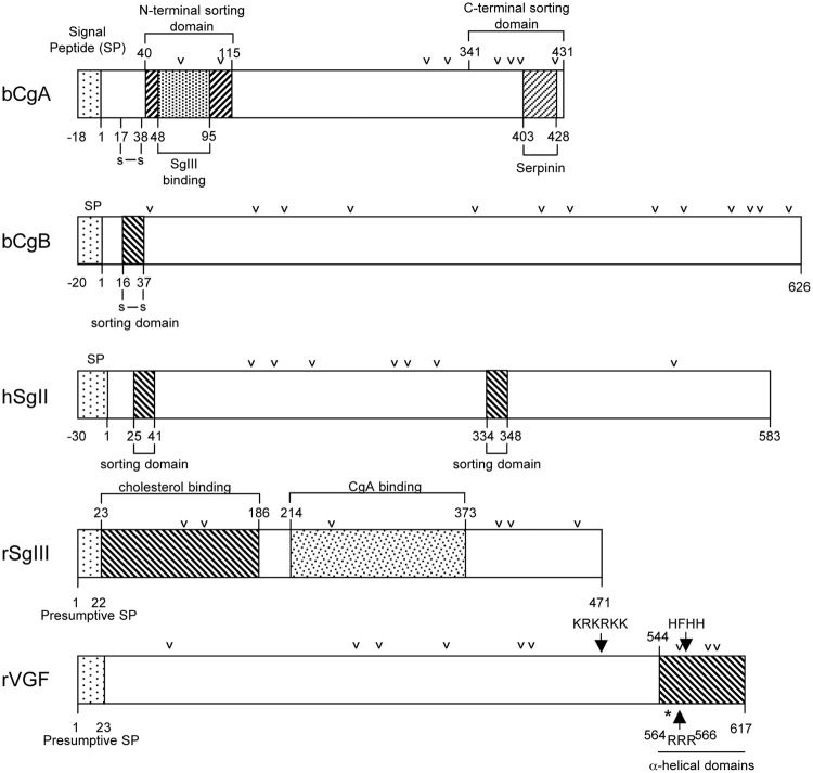
Fig. 6.
Schematic diagram showing the structures and sorting domains of bovine (b) CgA, bovine CgB, human (h) SgII, rat (r) SgIII, and rat VGF. The sorting determinants of these granins and the binding sites referred to in the text are indicated. The arrowheads represent paired or multiple basic residues that are potential PC cleavage sites. The asterisk in the VGF structure represents a noncanonical cleavage site 553RPR555 that is cleaved to generate a number of peptides.

