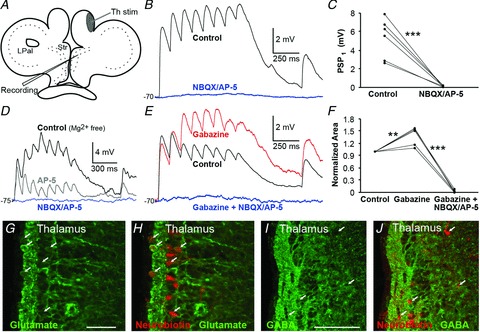
Figure 5. Thalamostriatal synaptic responses are glutamatergic.

A, schematic drawing indicating stimulation area of Th fibres in the most lateral region of the medial pallium. B, striatal postsynaptic potentials (PSPs) in regular aCSF in response to stimulation of Th fibres (black trace) and application of NBQX (40 μm) and AP-5 (50 μm), that completely removed all responses (blue trace), quantified by comparing the amplitude of the first PSP before and after drug application (C). D, NMDA and AMPA receptors were investigated in Mg2+-free aCSF by current-clamp recordings of striatal PSPs in response to stimulation of Th fibres (black trace), and after sequential application of AP-5 (grey trace) and both AP-5 and NBQX (blue trace) at about −75 mV. E, application of gabazine (20 μm, red trace) increased responses in recorded neurons (rest Vm−70 mV), indicative of oligosynaptic inhibition. Responses were completely removed by further application of NBQX and AP-5 (blue trace). F, quantification of the effect of drugs in (E). G, glutamate immunostaining (green) of the Th showed that the cell layer is packed with glutamatergic neurons. H, retrogradely labelled neurons (red) from the striatum are glutamatergic as indicated by the arrows and the co-staining. I, immunostaining for GABA (green) in the Th. J, retrogradely labelled neurons (red) were GABA-immunonegative as there was no co-staining with GABA. Scale bars, 50 μm in G; 100 μm in I. **P < 0.01, ***P < 0.001, two-tailed paired t tests. LPal, lateral pallium; Str, striatum; Th, thalamus.
