Fig 1.
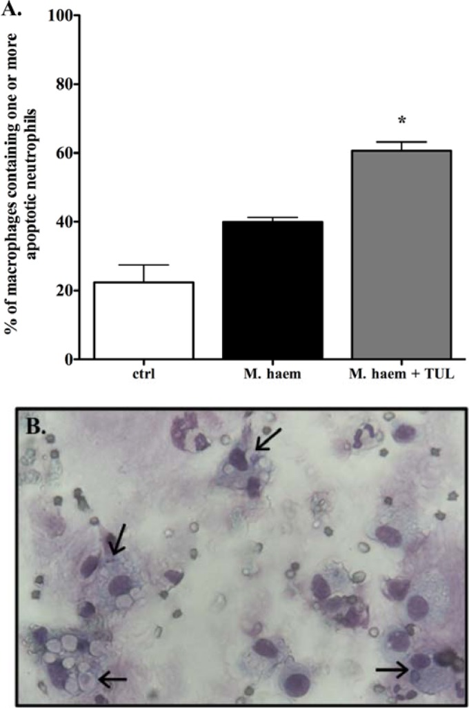
Alveolar macrophages readily phagocytose apoptotic neutrophils in tulathromycin-treated calves challenged with live M. haemolytica. Counts (A) and representative light microscopy image (B) of cytospin preparations of BAL fluid samples isolated from uninfected control calves (ctrl), sham-treated M. haemolytica-challenged calves (M. haem), and tulathromycin-treated M. haemolytica-challenged calves (M. haem + TUL) 3 h postinfection, stained with Diff-Quik and analyzed using light microscopy. Macrophages containing one of more apoptotic neutrophils were enumerated. Values were expressed as a percentage of total macrophages on each slide preparation. Values are means ± SEM. n = 3 to 7/group. *, P < 0.001 versus the control group. A minimum of 100 macrophages were counted in each sample for all treatment groups.

