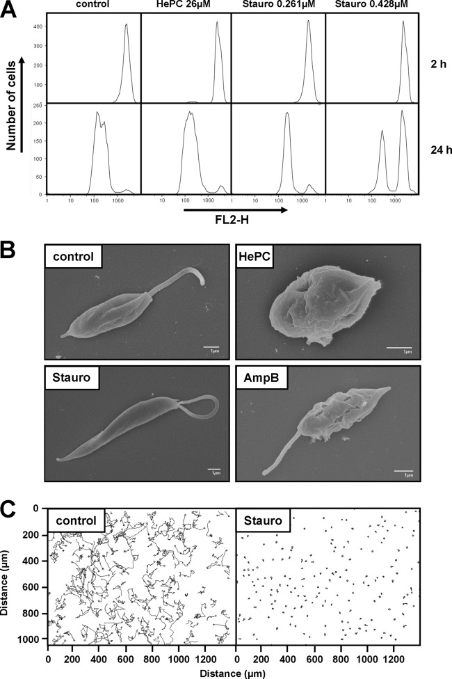
Fig 2.
Staurosporine treatment affects promastigote proliferation, morphology, and motility. (A) Proliferation assay of L. donovani in the presence of 26 μM miltefosine (hexadecylphosphocholine [HePC]) or 0.261 μM and 0.428 μM staurosporine (Stauro). L. donovani promastigotes from a logarithmic culture were stained with CFSE, and fluorescence levels were determined by FACS analysis using the fluorescence channel FL2-H at 2 h (top) and 24 h (bottom) after inoculation into new culture medium. A representative experiment of two independent tests is shown. (B) Scanning electron microscopy (SEM). L. donovani promastigotes from the logarithmic growth phase were treated for 4 h with miltefosine (HePC, 46 μM), staurosporine (Stauro, 0.428 μM), and amphotericin B (AmpB, 0.055 μM) and processed for SEM as described in Materials and Methods. The bar corresponds to 1 μm. Control corresponds to untreated promastigotes from a logarithmic culture. (C) Motility assay. The movement of L. donovani promastigotes in untreated and staurosporine-treated cultures was assessed by taking 200 pictures in 20 s using a phase-contrast microscope. Images were analyzed using the image analysis medeaLAB tracking software (version 5.5). Each line represents the path taken by a single L. donovani promastigote over the 20 s of the analysis.

