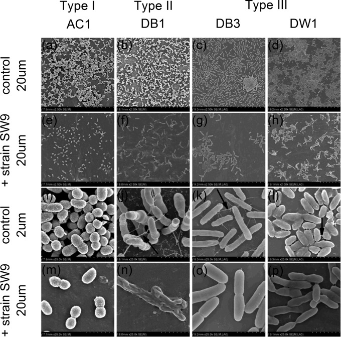
Fig 4.
SEM micrographs of biofilms developed by biofilm-forming strains with or without Bacillus sp. SW9 on PVC surfaces. (a to d) Dense biofilms developed by the biofilm-forming bacteria; scale bar, 20 μm. (e to h) Loose biofilms developed by the coculture of biofilm-forming bacteria and strain SW9; scale bar, 20 μm. (i to l) Cells were well connected with filaments (indicated by arrows); scale bar, 2 μm. (m to p) Cells were tightly bound with extracellular matrix or loosely connected with fewer filaments; scale bar, 2 μm.

