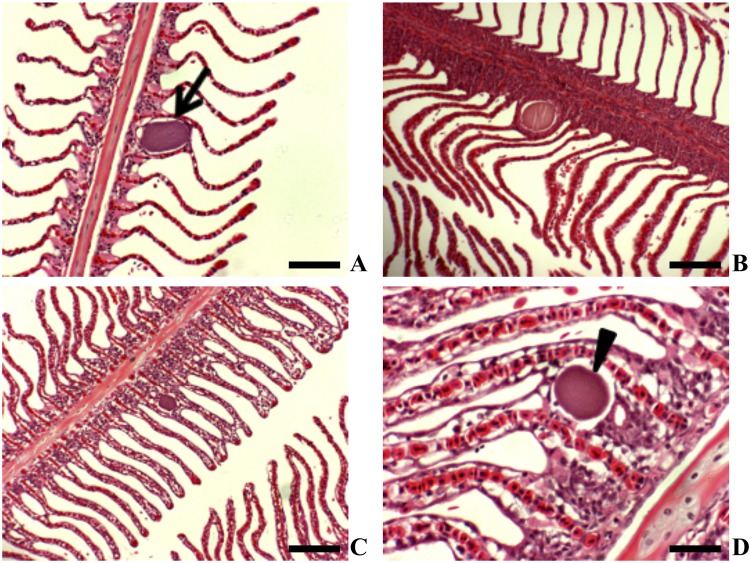
Fig 1.
Yellowtail kingfish gills showing epitheliocystis (H&E staining). (A) Basophilic inclusion with no associated host response (scale bar = 100 μm) (2008 cohort). (B) Membrane-enclosed inclusion at base of lamellae (scale bar = 100 μm) (2010 cohort). (C) Basophilic inclusion with an associated lamellar fusion (scale bar = 200 μm) (2008 cohort). (D) Higher magnification of basophilic inclusion at base of lamella associated with proliferation and vacuolation of the basal lamellar epithelium (scale bar = 50 μm) (2009 cohort).

