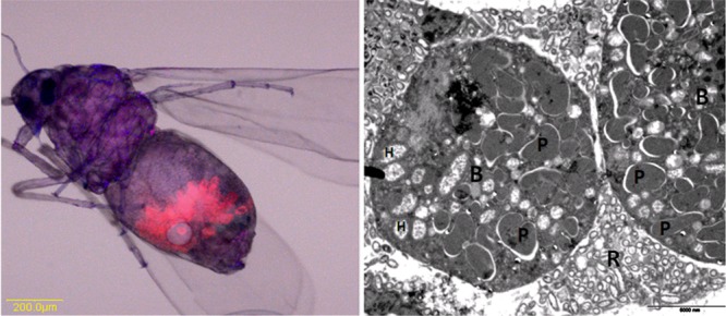
Fig 1.

(Left panel) “Ca. Portiera” localization in adult female B. tabaci by fluorescence in situ hybridization analysis with a probe specific for “Ca. Portiera” (red). Note that “Ca. Portiera” is restricted to bacteriocyte cells in the insect abdomen. (Right panel) transmission electron microscopic image showing two bacteriocytes (B) full of “Ca. Portiera” (P) and some cells of the secondary endosymbiont Hamiltonella (H) and surrounded by the secondary symbiont Rickettsia (R), which is scattered in the hemolymph. The bar in the right panel is 5 μm.
